
|
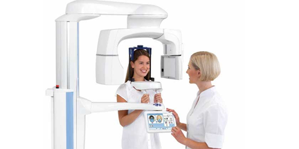
Accurate diagnosis and treatments are vital in dentistry and for this reason the cone beam computed tomography (CBCT) is dominating the industry. Its three-dimensional image viewing, and localising capabilities make it a firm favourite amongst dental clinicians. Unlike traditional CT scans, CBCT imaging can highlight dental structures, soft tissues, nerve paths and bone in the craniofacial region all from one single scan. 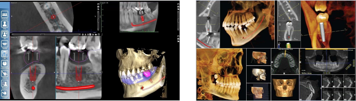 What is CBCT Cone Beam Computed Tomography (CBCT) systems are the latest in digital dentistry and are a variation on the traditional CT systems. Unlike traditional CT scans where “slices” are typically taken, the CBCT system has a cone-shaped x-ray beam which rotates around the patient and captures the data. CBCT scans are mainly used to assess the position of teeth, jaw joints, bone structure and the airway.
The reduction in radiation dosage occurs due to intelligent low dose 3D imaging algorithms which see dentists adhering to ALARA principles (As Low As Reasonably Achievable). Although Planmeca has significantly reduced radiation exposure, they have not compromised on the image quality. The ProMax is suitable for various clinical cases including orthodontics, implant planning, maxillofacial surgery, airways, and sinus studies. CALM Artifacts caused by patient movements are one of the biggest issues surrounding CBCT imaging. Not only do the artifacts impact the quality of the image but they also prolong the x-ray process. Planmeca have tackled such issues by introducing a software known as CALM. This software can be applied either before or after the scan to ensure patient movements do not impact the results. The CALM software reduces the number of retakes for dentists and enables the clinician to succeed every time. You can learn more about Planmeca’s CALM technology here: Planmeca CALM – New algorithm for patient movement correction Benefits A major advantage to CBCT imaging is the quality and accuracy of the images produced. The cone beam scan can zoom into specific areas from different angles allowing dentists to further examine key areas like single tooth roots. The versatility of digital imaging allows for a more accurate diagnosis. Due to the advancement in technology the cone beam CT can easily scan both bones and soft tissue, which enables the dentist to create a more precise treatment plan. Another advantage of CBCT imaging is that the process is very quick and painless. Typically, with a CT scan it takes between 20-40 minutes for the scan to be complete whereas with a CBCT scan the full mouth can be examined within just a few minutes. A dentist is only required to spend seconds on a specific area to get their result. A single scan produces hundreds of images that are from different views and angles, this enables the clinician to get a full view and evaluation of the patient's mouth. An additional benefit to this is the CBCT scan provides a non-invasive yet accurate, painless experience for the patient. Clinical benefits CBCT imaging provides several clinical benefits in dentistry, particularly in Implantology planning. These advanced scans allow dentists to measure and localise the available jawbone, meaning they can precisely do a virtual implant placement. With a CBCT and optical scan, Clinicians can produce a model of the patient's soft tissues, bones, and teeth in order to design the patient a bite. The advancements in the CBCT scan significantly reduces the risk of an implant being misaligned. This type of scan enables safer and predictable treatments, for instance; the chance of nerve damage occurring during an implant has been greatly reduced due to cone beam scans, this is because dentists now are now able to determine where the sensory nerves are and can carefully select the right implant length based on this. This is not achievable with a regular CT scan. Through CBCT imaging dentists are able to pinpoint the exact location of the impacted tooth which not only reduces surgery time, but it also facilitates post-operative recovery. In terms of endodontics the sub millimetric resolution produced by the CBCT image, shows up details with great precision. The advanced technology means 100% of apical lesions are detected compared to just 28% with a traditional x-ray. The cone beam has a significant clinical benefit on the diagnosis, treatment, and evaluation of maxillary sinus. It helps provide clarification of the carotid and other arteries which cannot be picked up on a traditional CT x-ray. The definitive benefit of the CBCT is that it allows clinicians to illustrate the implant treatment plans to patients, giving them greater understanding of the treatment options available to them. 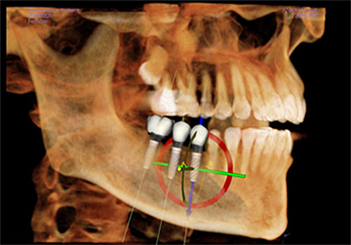 CBCT Implant Image Other than dental treatment planning, CBCT can also be used for advanced and complex cases such as;
With the shift from traditional dentistry to digital, the Cone Beam CT is fast becoming more and more popular in practices. If you want to learn more about 3D imaging visit: http://www.dentaldynamix.co.uk/promax-3d.html or contact our office to arrange a demonstration. As a thank you for reading our blog, we're giving away the below 'Atlas of Cone Beam' book to the first five readers. Contact us to receive yours. 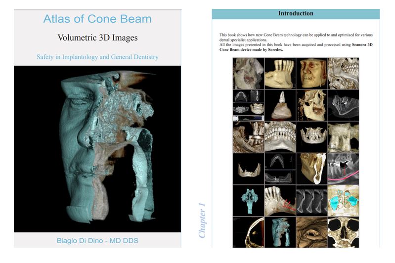
1 Comment
|
AuthorDental Dynamix ArchivesCategories |
 RSS Feed
|
We like to socialise!
|
|
©Dental Dynamix Imaging LTD, 07623713